


What is a macular hole?
The macula is the small central area of the retina. It provides the fine detail needed for reading, driving and recognising faces. A macular hole is a rupture through the full thickness of this tissue. Light entering the eye no longer lands on healthy retinal cells, so a blurred or blank patch appears in the middle of your vision.
Macular holes are uncommon. Population studies suggest they affect roughly 3 in every 1,000 people aged over 55. Around two thirds of cases occur in women, and most patients are between 60 and 80 years old. The condition is painless and affects one eye first in most people.
What are the symptoms of a macular hole?
Symptoms usually develop gradually over weeks or months. They affect central vision only. Peripheral vision is preserved, so people rarely lose their way around.
- Distorted or wavy central vision
- A blurred or blank patch in the centre of vision
- Difficulty reading or recognising faces
- Straight lines, such as door frames, appearing bent
- Objects appearing smaller or further away with the affected eye
Many people notice the problem only when they cover the healthy eye. If you have sudden distortion, new floaters or flashing lights, seek an urgent eye assessment. These symptoms can also signal a retinal tear or detachment. The NHS conditions guidance explains when eye symptoms need same day review.
What causes a macular hole?
Most macular holes are age related. The vitreous gel that fills the eye shrinks over time and separates from the retina. If the gel remains stuck to the macula, it tugs on the delicate tissue until a hole forms. This process is called vitreomacular traction.
- Age related shrinkage of the vitreous gel, the most common cause
- Blunt injury to the eye
- Long standing swelling of the macula, for example after retinal vein occlusion or diabetic disease
- High myopia, which stretches the retina
- Previous retinal detachment surgery
About one in ten patients later develops a hole in the second eye, so both eyes are monitored. Conditions causing macular swelling are covered further on our medical retinal conditions page.
How is a macular hole diagnosed?
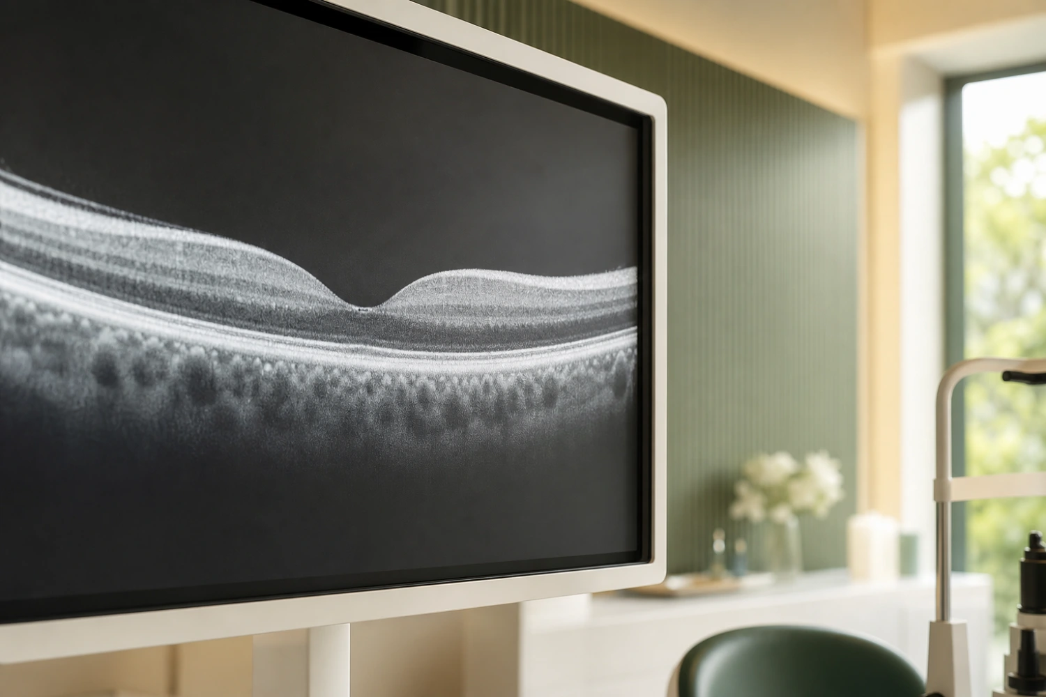
Diagnosis is straightforward with modern imaging. Optical coherence tomography (OCT) produces a cross sectional scan of the retina in seconds. It confirms the hole, measures its width and shows whether the vitreous is still pulling on the macula.
Your assessment with Mr Zine Elhousseini typically includes a vision test, dilated examination of the retina and an OCT scan. Hole size guides treatment, because smaller holes generally close more readily than larger, long standing ones. Early referral therefore matters.
How it's treated
Macular holes are repaired surgically through vitrectomy, where the vitreous gel is removed and the hole is closed, often with the help of a gas bubble to let the retina settle back into place. Most patients notice a steady improvement over the following weeks.
Quick facts
Treatment
Vitrectomy
Time
45 to 90 mins
Recovery
1 to 4 weeks
Anaesthetic
Local or general
Related treatments
Concerned about your vision?
Book a consultation with Mr Elhousseini for a clear diagnosis.